A worldwide community photographing and learning about wildlife

Project Noah Nature School

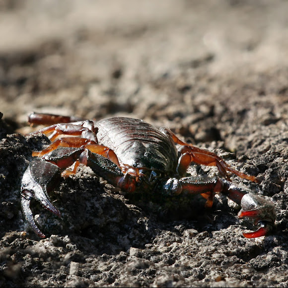
Euscorpius italicus (Herbst, 1800)
The fine structure of scorpion sensory organs. I. Tarsal sensilla Rainer F. Foelix and Joachim Schabronath Ruhr Universitat Bochum, Lehrstuhl fur Zellphysiologie, . Postfach 102 148, D 4630 Bochum, West Germany Summary The fine structure of the following sensory organs on the tarsi and basitarsi of scorpion legs was investigated: (1) mechanoreceptive hairs and bristles, (2) slit sensilla, (3) chemoreceptive hairs, and (4) tarsal organs. The mechanoreceptive hair sensilla are innervated by seven bipolar neurons, the dendrites of which are attached to the hair base. A large multicellular gland underlies the hair socket. The slit sensilla have two neurons each. The first neuron gives off a single dendrite which attaches to the cover membrane of the slit. The second neuron gives rise to two dendrites which penetrate the slit but do not reach the cover membrane. The chemoreceptive hairs are associated with 22-23 neurons; four dendrites end as mechanoreceptive dendrites at the hair base, the other dendrites enter the double lumen of the hair shaft and become exposed to the outside through a pore at the hair tip. The tarsal organ, situated dorsally near the distal end of the tarsus, is a small pit with two pores inside. Six to nine dendrites are exposed at each pore, which implies a chemoreceptive function (olfaction?). The structure of the various receptors closely resembles that of spider sensilla. In comparison with insect sensory organs, scorpion and spider sensilla are exceptional in always having multiinnervated mechanoreceptive hairs and a higher number of sensory cells per sensillum. https://www.flickr.com/photos/morton1905...

Spotted on Apr 10, 2018
Submitted on Apr 10, 2018


No Comments