A worldwide community photographing and learning about wildlife

Project Noah Nature School

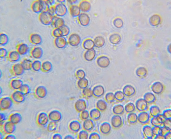
Didymium sp (in progress) possibly Nigripes spores
Brownish violet, minutely spinulose or nearly smooth, 8-10 µm diam ( http://hiddenforest.co.nz/slime/family/p... ) . ( sporangium ---> http://www.projectnoah.org/spottings/795... ) - ===My microscope magnified till x1200, but for this pictures at the microscope i have to take 1 ocular out and replace it with the usb-microscope-camera. The camera therefore cost me one magnification and some quality of the pictures (Picture magnification at ca x600-x800 !! [i suspect- i have still to work that out !]. --- I saw no remarkable/eyecatching spines, the surface seems for me smooth!
The Amoebozoa are a major group of amoeboid protozoa, including the majority that move by means of internal cytoplasmic flow. Their pseudopodia are characteristically blunt and finger-like, called lobopodia. Most are unicellular, and are common in soils and aquatic habitats, with some found as symbiotes of other organisms, including several pathogens. The Amoebozoa also include the slime moulds, multinucleate or multicellular forms that produce spores and are usually visible to the unaided eye. Amoebozoa vary greatly in size. Many are only 10-20 μm in size, but they also include many of the larger protozoa. The famous species Amoeba proteus may reach 800 μm in length, and partly on account of its size is often studied as a representative cell. Multinucleate amoebae like Chaos and Pelomyxa may be several millimetres in length, and some slime moulds cover several square feet ( http://www.hiddenforest.co.nz/slime/inde... ), ( http://www.discoverlife.org/mp/20q?searc... ), ( http://slimemold.uark.edu/fungi/default.... )
The Didymiaceae family, like all members of the Physarales order are distinguished by the presence of crystalline lime. Which is restricted to the peridium in the form of crystals or disks. Spores are typically dark brown to dark purple-brown. ------Genus: Didymium Sporangia stalked, sessile, or forming plasmodiocarps; sporangia wall membranous or cartilaginous, with superficial crystals of lime either scattered over the surface or combined into a separable crust. Capillitium of branching threads, which are thickened at intervals with dark calyciform nodes, in normal developments without lime. --------- (i think those glassy stripes,could be the lime [my first microscope since years, so i will surely make enough mistakes!!].

Spotted on Dec 25, 2011
Submitted on Dec 28, 2011


2 Comments
I love these microscopic shots.
Nice! Love to see microscope pics in Noah! :-)