A worldwide community photographing and learning about wildlife

Project Noah Nature School

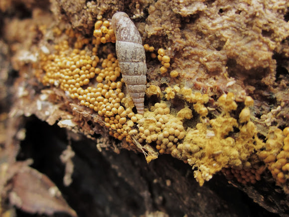
Trichia favoginea var. persimilis (P. Karst.) Y. Yamam. 1998
Sporocarps sessile, clustered, subglobose, 0.5-0.8 mm diam., shining ochraceous to yellow-brown, seated on a common, yellow-brown hypothallus. Peridium membranous, shining, the inside marked with rows of warts which are often arranged in lines. Capillitium of ochraceous-yellow elaters, 4-6 µm diam., marked with 4-5 closely-set spiral bands and studded with short spines, longitudinal striae inconspicuous. Spore-mass ochraceous yellow. Spores pale yellow, 11-14 µm diam., marked with a broken reticulation of small meshes, the non-reticulate areas covered with large warts, border incomplete in optical section. Plasmodium white ( http://www.discoverlife.org/mp/20q?searc... )
These colonies developed from an earlier plasmodial phase; at that stage, the slime mould really is slime (it looked like wallpaper paste). The plasmodium moves over the wood rather like a giant amoeba, ingesting bacteria. It later "fruits", forming the spore-producing structures (sporocarps/fruitingbodies) shown here; the sporocarps are the individual small rounded structures visible in the photo. What is left over is the so-called hypothallus, visible here as a translucent substance on picture #3 and a little at #2. ( http://www.geograph.org.uk/photo/1524494... ) ---------------------- dead wood,decaying wood, and other organic material,north america and europe, cosm.
( http://www.mycobank.org/MycoTaxo.aspx?Li... ), ( http://www.nederlandsesoorten.nl/nsr/con... )

Spotted on Mar 11, 2012
Submitted on Mar 13, 2012


2 Comments
Do you know maybe what is that cocoon on the 1st photo? I have the same spotting of that: http://www.projectnoah.org/spottings/919...
nice series showing all the stages